

Share this article
Researchers develop tool to detect amphibian disease
While Perkinsea may be less widely known than amphibian diseases such as chytrid fungus and ranavirus, the pathogen is also killing off frogs throughout the United States and around the world.
But researchers didn’t have a good way to detect the pathogen, which enters the frog, goes straight to the liver and spreads until the frog dies — until now.
“Perkinsea is very understudied right now,” said Emily Karwacki, lead author of the study published in Diseases of Aquatic Organisms and associate researcher at the University of Central Florida. “At the time, there were still a lot of scientists that didn’t think it was worth noting or studying. Within the past year or so, it’s becoming recognized as something that’s a cause for concern and should be studied more.”
Karwacki, who was an undergrad at the start of her research, worked with her adviser Dr. Anna Savage to develop a way to detect the pathogen in frogs. In the past, researchers used histopathology, which uses a microscope to view tissue and look for spores, to detect the pathogen, but it proved to be labor intensive and time consuming and requires killing the animal, Karwacki said.
Karwacki knew molecular biology methods, which she had already been working on in the lab, would be a good start. She used qPCR, or a quantitative polymerase chain reaction, which uses a short probe that fits inside two primers to determine if a particular gene is present. The probe attaches and emits fluorescent light, and a machine quantifies how much light is produced to determine whether and how much of the gene is present. In this study, Karwacki identified a gene sequence unique to amphibian Perkinsea and no other related organism, so that her qPCR test would be specific.
“It was exciting but also really difficult,” Karwacki said. “I started the project, and I knew it was going to be a tough one, but I also knew it was going to be worth it.”
Using qPCR, Karwacki and her colleagues sampled frogs at three sites in Florida, including Gold Head Branch State Park in Keystone Heights, the UCF Arboretum in Orlando and the Archbold Biological Station in Venus. Twenty-five percent of the frogs sampled tested positive for Perkinsea, and frogs farther north tested positive in higher numbers.
Karwacki hopes this method of detecting Perkinsea can help figure out where the pathogen has reached and how long it has been around. So far, she and her team have received requests from around the country and beyond for the paper and for details of implementing their methodologies.
Karwacki is currently working on sampling tissues in museum specimens that go back at least 100 years to help determine how long the pathogen has been around in certain areas and which species might be a reservoir host.
“I’m hoping studies like that could be utilized all over the world,” she said.

Header Image: Karwacki developed a method to screen frogs for the infectious disease caused by the Perkinsea pathogen. The disease is linked to mass die-offs of frogs around the world. ©University of Central Florida
Related Posts
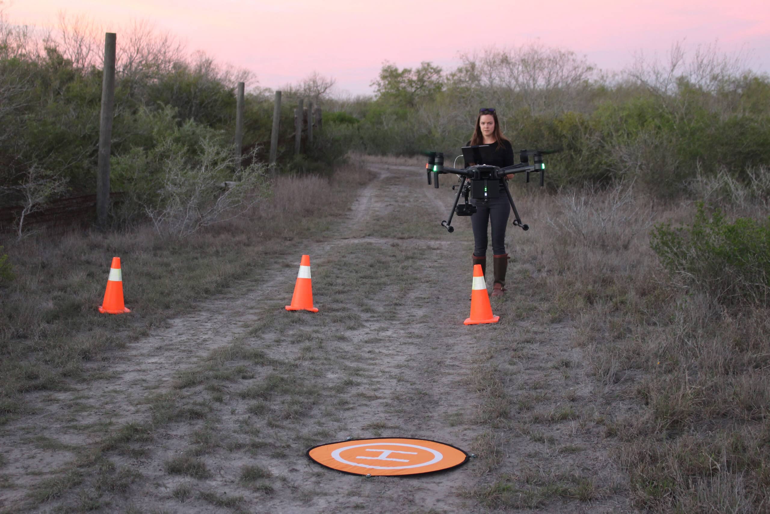
WSB: Study tests accuracy of thermal drone surveys
In South Texas, the heat can be blinding
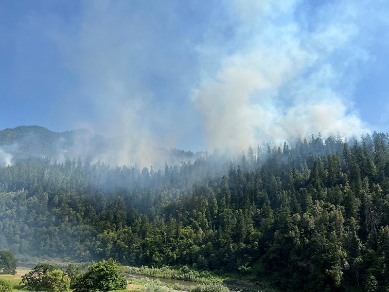
Computer model explores Tribal use of fire for ecosystem health
The Karuk Tribe regularly conducted burns in the fire-prone Klamath Mountains

2024 TWS Elections: Southwest Representative
This year’s nominees for Southwest Representative to TWS Council are Kathy Granillo and Erika Nowak